Left Hip Muscles Anatomy / Muscles Of The Hips And Thighs Human Anatomy And Physiology Lab Bsb 141 - Learn their anatomy efficiently and easily using kenhub's muscle anatomy and reference charts!
Left Hip Muscles Anatomy / Muscles Of The Hips And Thighs Human Anatomy And Physiology Lab Bsb 141 - Learn their anatomy efficiently and easily using kenhub's muscle anatomy and reference charts!. One example of an ab exercise that actually focuses. We study anatomy at the practical anatomy class we study the human body. There are a lot of muscles of the hip and thigh. The muscles and the bones are under the layer of subcutaneous fat. A radiograph is not as helpful in diagnosing trochanteric bursitis as soft tissues and muscles are not visible to any degree(15).
Attached to the bones of the skeletal system are about 700 named. The hip muscles encompass many muscles of the hip and thigh whose main function is to act on the thigh at the hip joint and stabilize the pelvis. These muscles constitute the anatomical classification known as the medial compartment of the thigh. This webpage presents the anatomical structures found on hip mri. This muscle assists with the external rotation of the hip.
Muscles Of The Leg And Foot Classic Human Anatomy In Motion The Artist S Guide To The Dynamics Of Figure Drawing from doctorlib.info This anatomical atlas was especially designed for a specific public (radiologists, surgeons, rheumatologists and physicians specializing in musculoskeletal imaging). Pick which works for you and then. Anatomy 3d atlas allows you to study human anatomy in an easy and interactive way. Muscle movements, types, and names. The hip's unique anatomy enables it to be both extremely strong and amazingly flexible, so it can bear weight and allow for a wide range of movement. The geometry of the hip allows wide range of motion in all planes. Leave a reply cancel reply. Comprehensive information about hip joint anatomy including muscles, tendons, ligaments, bones, bursae, skeletal structure and joint capsules.
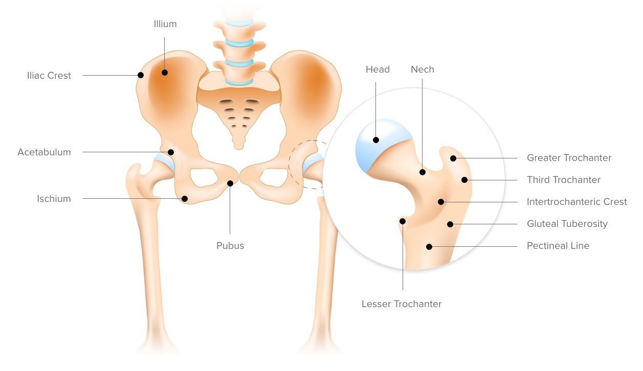
The hip bone, also known as the innominate bone, coxal bone or os coxae, is a large bone that sits in the pelvis.
If left unstretched, shortened hip flexors affect the position of the pelvis, which in turn affects the position and movement of the lower back. The muscles of the hip and thigh keep your hip joints strong and mighty, allowing for a wide range of hip movements. One example of an ab exercise that actually focuses. The muscles and the bones are under the layer of subcutaneous fat. The main functions of the neck muscles are to permit movements of the neck or head and to provide structural support of the head. Learn their anatomy efficiently and easily using kenhub's muscle anatomy and reference charts! In human anatomy, the muscles of the hip joint are those muscles that cause movement in the hip. Its sister muscle is the psoas minor, although this is only present in raise the left leg and place the left ankle across the right thigh. Comprehensive information about hip joint anatomy including muscles, tendons, ligaments, bones, bursae, skeletal structure and joint capsules. We study anatomy at the practical anatomy class we study the human body. Several muscles cross the front of the hip and create hip flexion, pulling the thigh and trunk toward each other, but probably the most important is the iliopsoas. A radiograph is not as helpful in diagnosing trochanteric bursitis as soft tissues and muscles are not visible to any degree(15). Through a simple and intuitive interface it is possible to observe every anatomical structure from any angle.
The main functions of the neck muscles are to permit movements of the neck or head and to provide structural support of the head. for detailed anatomy of pelvic bones, read anatomy of hip bone. It is a flat, triangular muscle on the anterior wall of the pelvis. Highly detailed 3d models, with textures up to 4k resolution, enable to examine the shape of each. The muscles of the neck can be divided into groups according to their location.
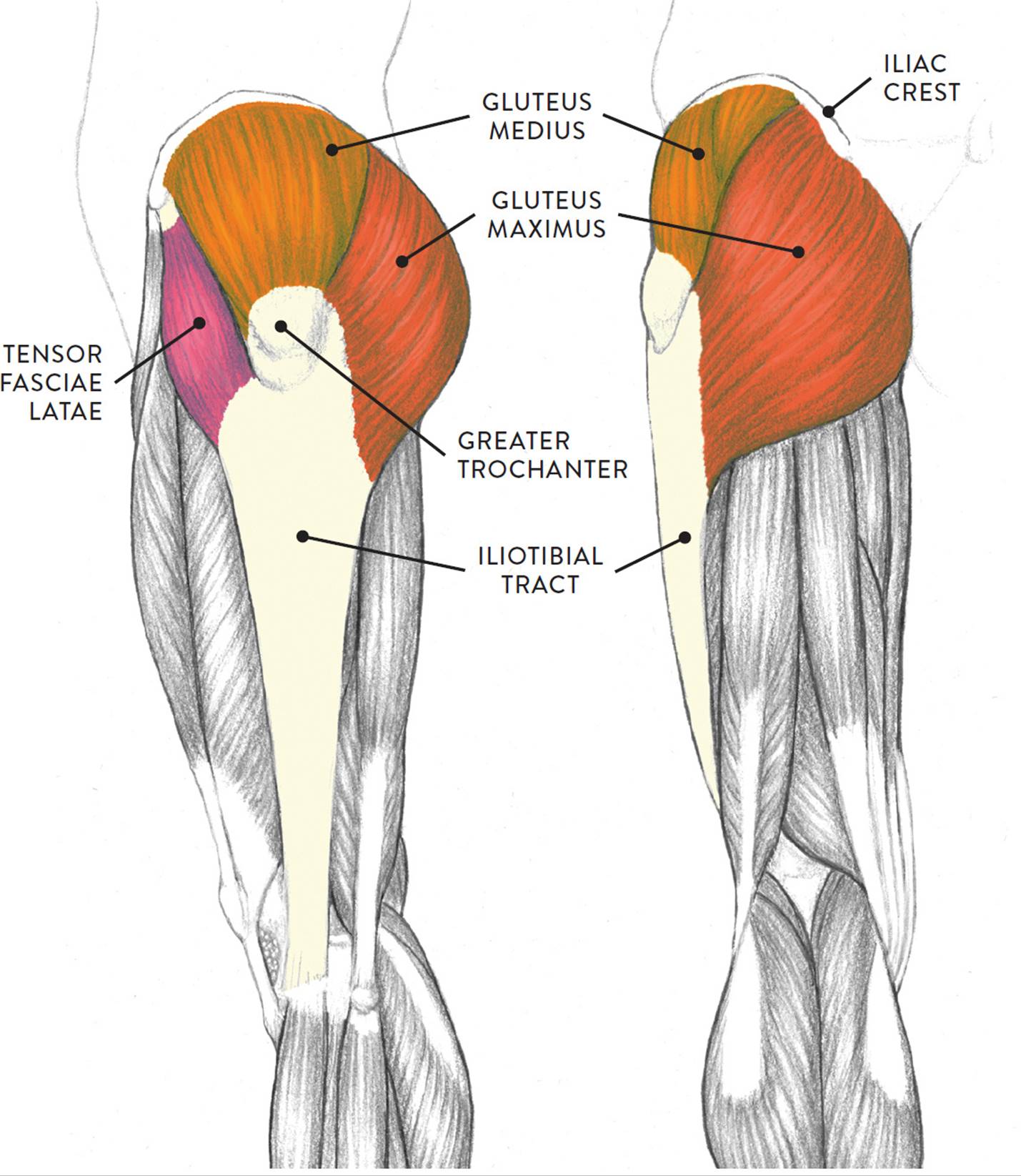
Tendinitis And Bursitis Treatment Cincinnati Tendinitis Dayton Oh from www.beaconortho.com We study anatomy at the practical anatomy class we study the human body. Groin, inguinal region and the anterior. Pelvis and acetabulum, with muscle attachment sites. The hip joint is an intricate structure including hip bones, hip articular cartilage, muscles, ligaments and tendons, and synovial fluid. There are a lot of muscles of the hip and thigh. Learn their anatomy efficiently and easily using kenhub's muscle anatomy and reference charts! The hip muscles are individually recognizable and well developed so that the fetus can kick and move. Attached to the bones of the skeletal system are about 700 named.
These muscles constitute the anatomical classification known as the medial compartment of the thigh.
I pulled some muscles on left hip hiking. This muscle assists with the external rotation of the hip. If left unstretched, shortened hip flexors affect the position of the pelvis, which in turn affects the position and movement of the lower back. It is a flat, triangular muscle on the anterior wall of the pelvis. Anatomy of the muscular system. Learn their anatomy efficiently and easily using kenhub's muscle anatomy and reference charts! Muscles of the hips and thighs | human anatomy and. The muscles and the bones are under the layer of subcutaneous fat. Muscles that act on the lower limb cause movement at the hip, knee and foot joints. Muscle movements, types, and names. We study anatomy at the practical anatomy class we study the human body. 3 months later i got acute excrutiating pain in inguinal area. Your email address will not be published.
Attached to the bones of the skeletal system are about 700 named. Diarthrodial joint with its inherent stability dictated primarily by its osseous components/articulations. These muscles constitute the anatomical classification known as the medial compartment of the thigh. The hip joint is an intricate structure including hip bones, hip articular cartilage, muscles, ligaments and tendons, and synovial fluid. In order to isolate the abdominals, you need to minimize the involvement of the hip flexors and maximize the contraction of the abdominals.
Everything You Need To Know About Lateral Hip Pain The Physio Depot from 44wj5q2j6wo23s4mp6owjohh-wpengine.netdna-ssl.com The hip is a complicated mechanism and therefore hip pain can originate in many different parts of the joint. This webpage presents the anatomical structures found on hip mri. How many of the 11 muscles involved in hip flexion can you name from memory? 3 months later i got acute excrutiating pain in inguinal area. The hip joint is an intricate structure including hip bones, hip articular cartilage, muscles, ligaments and tendons, and synovial fluid. The different anatomical areas of the gluteal region: Attached to the bones of the skeletal system are about 700 named. Through a simple and intuitive interface it is possible to observe every anatomical structure from any angle.
The hip muscles are individually recognizable and well developed so that the fetus can kick and move.
Muscle movements, types, and names. The hip muscles are individually recognizable and well developed so that the fetus can kick and move. The hip's unique anatomy enables it to be both extremely strong and amazingly flexible, so it can bear weight and allow for a wide range of movement. The hip bone, also known as the innominate bone, coxal bone or os coxae, is a large bone that sits in the pelvis. A radiograph is not as helpful in diagnosing trochanteric bursitis as soft tissues and muscles are not visible to any degree(15). The main functions of the neck muscles are to permit movements of the neck or head and to provide structural support of the head. 936 x 504 png 317 кб. Groin, inguinal region and the anterior. The different anatomical areas of the gluteal region: The muscular system is responsible for the movement of the human body. Leave a reply cancel reply. Learning the anatomy of your hip will better enable you to pinpoint your pain and work with your doctor to keep it from limiting your life. In order to isolate the abdominals, you need to minimize the involvement of the hip flexors and maximize the contraction of the abdominals.


0 Komentar